
Preventive dentistry is an essential part of maintaining a healthy smile. In addition to professional cleanings and check-ups, preventive dentistry includes dental X-rays. These X-rays are pictures of the teeth, gums, and bones in the mouth and can help identify problems that might not otherwise be visible during a routine check-up. Many patients-- and particularly those who suffer from dental phobia-- have some degree of anxiety associated with getting dental X-rays. Learning more about the purpose of the X-rays and what to expect during the X-ray process can help alleviate this anxiety.
Why are dental X-rays necessary?
Dental X-rays are used for a variety of reasons, including:
- Early detection of problems. Dental X-rays can often detect problems early on, allowing for prompt treatment and prevention of progression. Problems detected early through X-rays include tooth decay, bone damage, and injuries like broken tooth roots.
- Identify impacted teeth. Sometimes, teeth are too crowded to break through the gums; this is known as impacted teeth. Dental X-rays help to identify this concern so that a treatment plan can be developed.
- Find permanent teeth. Dentists often use X-rays to determine where permanent teeth are located when children still have their baby teeth.
- Treatment planning. When certain procedures are required, dental X-rays can help dentists plan for the upcoming treatment. For example, dentists often consult X-rays to help plan for root canal surgery, treatment of large cavities, and implant placement.
- Identify abnormalities. If there are any abnormalities present-- including cysts, tumors, or abscesses-- dental X-rays help to determine the exact location so that a treatment plan can be developed.
What types of X-rays do dentists use?
The most commonly used X-rays include:
- Bitewing. These X-rays display the upper and lower back teeth. Dentists typically use them to check for decay between the teeth and to determine how the upper and lower teeth line up.
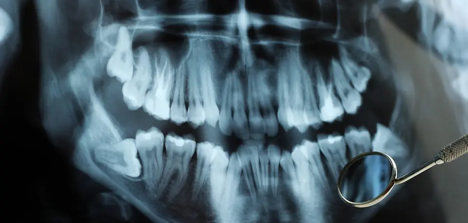
- Panoramic. Panoramic X-rays are not for cavity detection. Instead, they check for impacted teeth, injuries, and abnormalities like tumors, growths, and cysts. These X-rays allow the dentist to view a large area, including the teeth, jaws, sinuses, and temporomandibular joints.
- Digital. These X-rays use significantly less radiation than traditional dental X-rays. A sensor is placed in the mouth to capture images of the teeth; these images can then be sent to a computer, where they are typically saved.
- Occlusal. Occlusal X-rays allow the dentist to view the roof or floor of the mouth. This can help the dentist identify cleft palate, fractures, extra teeth that haven't broken through the gums, and other abnormalities.
- Periapical. These X-rays give a comprehensive view of the entire tooth-- from crown to root and even the surrounding bones. Because of this, problems that occur below the gum line can be identified.
What is the X-ray process like?
The dental X-ray process is simple and occurs right in your dentist's office. There is nothing you need to do to prepare ahead of time. However, if you are pregnant or suspect that you might be pregnant, it's important to disclose this information to your dentist prior to the X-ray process.
To begin the X-ray process, you will first have a lead apron placed over your body. You will then be asked to bite down on a plastic piece that holds the X-ray film. You will likely have to repeat this process a few times to ensure that the dentist gets all of the necessary views of your teeth. Alternatively, if your dentist uses digital radiography, an electronic sensor is used in place of traditional X-ray film. Upon completion, your dentist will read the X-rays and discuss the results with you.
For additional questions or concerns regarding dental X-rays, contact us today.